
• Cart
    • Quote
    • Inquiry
    • Cart
    • Quote
    • Inquiry
Novogene
  • Novogene
  • Genomics
    • Human Whole Genome Sequencing
    • Whole Exome Sequencing
    • Plant and Animal Whole Genome Sequencing
    • Microbial Whole Genome Sequencing

    Metagenomics

    • Shotgun Metagenomics Sequencing
    • Amplicon Sequencing

    Transcriptomics

    • mRNA Sequencing
    • Swift & Express mRNA Sequencing New!
    • Full-Length Transcriptome Sequencing
    • Prokaryotic RNA Sequencing
    • Metatranscriptome Sequencing
    • Total RNA Sequencing
    • Small RNA Sequencing (sRNA‑seq)
    • Whole Transcriptome Sequencing

    Single Cell & Spatial Omics

    • 10x Single Cell Gene Expression
    • Illumina PIP-seq Single Cell 3’ RNA Sequencing New!
    • Spatial Transcriptomics Sequencing New!

    Epigenomics

    • Whole Genome Bisulfite Sequencing (WGBS)
    • Enzymatic Methylation Sequencing
    • Directed Methylation Sequencing (DM-Seq) New!
    • RNA Immunoprecipitation Sequencing (RIP-seq)
    • Chromatin Immunoprecipitation Sequencing (ChIP-seq)
    • Cleavage Under Targets & Tagmentation (CUT&Tag) New!
    • Assay for Transposase-Accessible Chromatin with Sequencing (ATAC-seq)
    • Reduced Representation Bisulfite Sequencing (RRBS)

    Proteomics

    • Quantitative Proteomics New!
    • PTM Proteomics New!
    • Olink Proteomics New!

    Metabolomics

    • Untargeted Metabolomics

    Premade Library

    • Sequencing Only on Illumina Sequencer
    • Sequencing Only on Ultima Sequencer
  • PromotionsPromotions
    • Platforms
    • Service & Support
    • Automated Delivery Platform (Falcon)
    • Bioinformatics Analysis Tool (NovoMagic)
    • Customer Service System (CSS)
    • Case Study
    • Blog
    • Webinar
    • Brochure
    • Cancer Research
    • Immuno-oncology
    • Agrigenomics
    • Environment
    • Food Science
    • Human Microbiome
    • Plant and Animal Microbiome
    • Drug Discovery and Development
    • Rare and Complex Diseases
    • About Us
    • Our Locations
    • News & Events
    • Careers
  • Contact UsContact Us
    • mRNA Sequencing
    • Illumina Lane Sequencing
  1. Home
  2. Resources
  3. Blog
  4. Mapping Cancer in Context with 10x Visium HD

Mapping Cancer in Context with 10x Visium HD
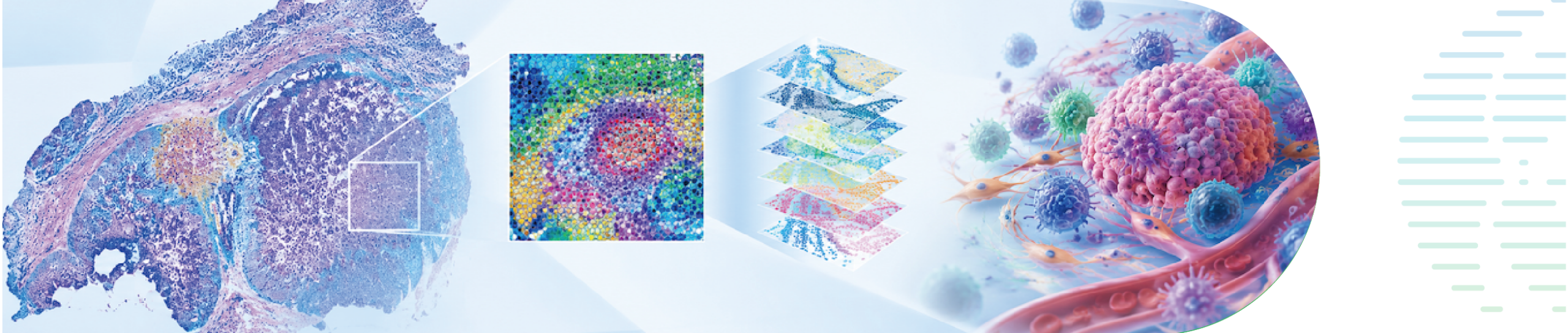
Cancer develops within a complex tissue environment. Tumor cells, immune cells, stromal cells, and blood vessels occupy different locations—and those spatial relationships can change as a tumor progresses or responds to treatment. Visium HD Spatial Gene Expression helps researchers examine gene expression alongside tissue morphology at single-cell scale resolution, making it possible to study cancer in its spatial context.

Five questions spatial oncology can answer

A spatial study often begins with a simple question: who is where? Mapping cell populations across a tissue section can reveal rare cell niches, the organization of tumor clones, and the distribution of immune cells around the tumor.

From there, researchers can ask:

· What are these cells doing? Identify gene expression programs associated with immune activation, exhaustion, invasion, or interactions between tumor and stromal cells.

· How do regions differ? Compare molecular and immune features across tumor cores, invasive margins, metastases, and other tissue regions.

· What changes over time or with treatment? Examine spatial patterns in paired samples collected before and after therapy, or across stages of tumor progression.

· What could be targeted or measured? Locate candidate biomarkers, cell populations, and pathways for further validation.

Key applications in cancer research

Tumor heterogeneity and evolution. Spatial profiling can show how tumor cell states and molecular programs vary within a lesion and between primary and metastatic sites. This helps researchers investigate clonal organization, invasive margins, and mechanisms associated with progression or drug resistance.

The tumor immune microenvironment. The location of an immune cell can be as informative as its identity. Researchers can examine immune infiltration, T and NK cell states, stromal barriers, and the organization of structures such as tertiary lymphoid structures. These patterns may help generate hypotheses about antitumor immunity and treatment response.

Biomarker and therapeutic target discovery. Spatially localized expression can help prioritize candidate markers that might be missed when a tissue is analyzed in bulk. Paired with clinical information and validation experiments, spatial data can support studies of treatment response, resistance, and patient stratification. It can also provide training data for models that connect pathology images with molecular features.

Designing a study around the biological question

The most useful spatial study starts with the comparison it needs to make. Examples include primary versus metastatic tumors, tumor versus adjacent tissue, early versus advanced disease, or samples collected before and after treatment. Depending on the question, researchers may combine spatial gene expression with single-cell RNA sequencing to refine cell-state definitions, then use pathology review, protein staining, or other assays to validate selected findings.

To translate these scientific questions into study designs, the figure below outlines seven cancer research directions, each with a suggested cohort, sample type, and biological question (Figure 1).
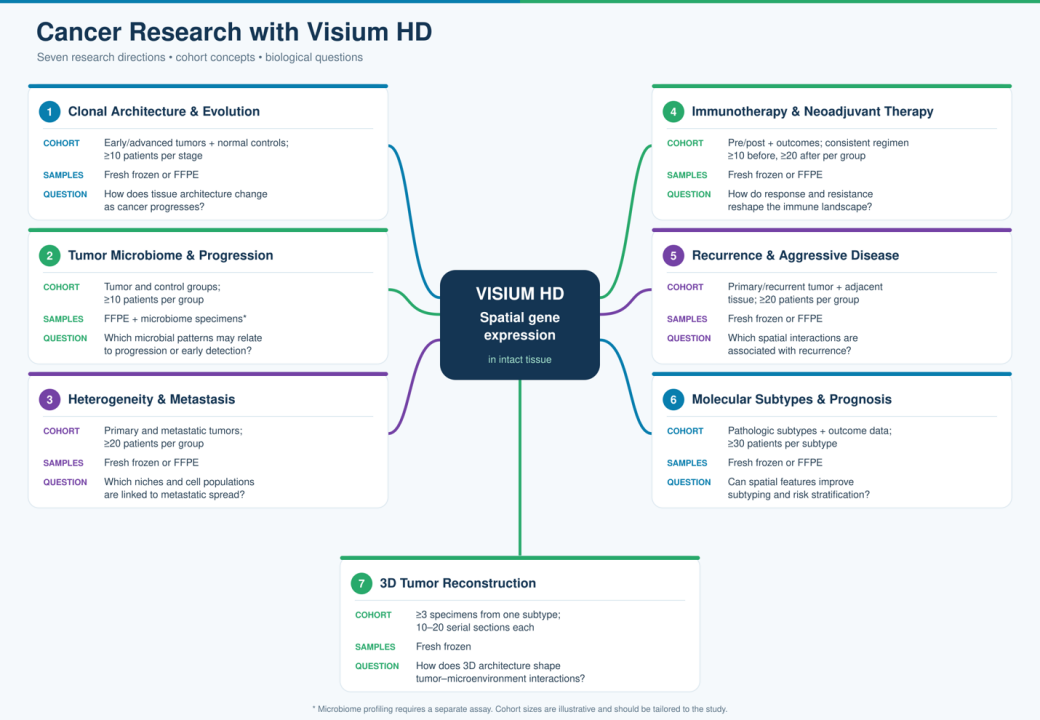
Figure 1. Seven illustrative Visium HD research directions in oncology, with example cohort designs, sample types, and biological questions.

Sample quality, tissue morphology, region selection, and the availability of matched clinical information should all be considered during study design. Visium HD supports FFPE, fresh frozen, and fixed frozen tissue sections, giving researchers options for both prospective collections and suitable archived material. Once samples are collected, the analysis workflow moves from data processing and spatial visualization to functional interpretation, with advanced and customized analyses selected according to the research question (Figure 2).
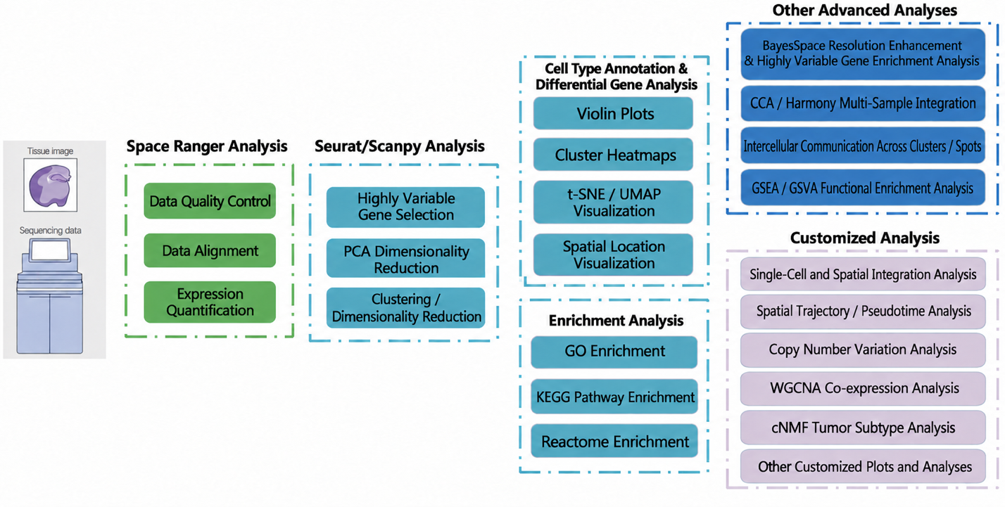
Figure 2 Bioinformatics data analysis workflow of Visium HD from SpaceRanger processing and Seurat/Scanpy analysis to spatial visualization, functional enrichment, and customized analysis.

Spatial research in action

In a Nature study of pancreatic cancer, researchers compared primary tumors with metastases in the liver, lung, and peritoneum. Their spatial maps revealed changes in cancer-cell states across sites, illustrating how metastatic tumors can differ even within one patient. Read the study.

In a Cell study of breast cancer, researchers trained Path2Space on spatial transcriptomics data to predict molecular features from pathology images. Applied to a larger tumor cohort, the model identified spatially defined microenvironment subgroups and features associated with treatment response. Read the study.

These studies demonstrate the breadth of questions spatial data can address. With Visium HD, researchers can build detailed tissue maps to investigate tumor organization, immune interactions, disease progression, and candidate biomarkers.

Planning a spatial oncology project? Contact Novogene to discuss your research question, sample type, and Visium HD study design.

ServicesServices menu

CompanyCompany menu

Contact UsContact Us menu

Service SupportService Support menu

Services
mRNA SequencingSwift & Express mRNA SequencingTotal RNA SequencingHuman Whole Genome SequencingWhole Exome Sequencing10x Single Cell Gene ExpressionIllumina PIP-seq Single Cell 3’ RNA SequencingSpatial Transcriptomics SequencingWhole Genome Bisulfite Sequencing (WGBS)Quantitative ProteomicsUntargeted MetabolomicsShotgun Metagenomics SequencingMetatranscriptome SequencingSequencing Only on Illumina SequencerSequencing Only on Ultima SequencerFull-Length Transcriptome SequencingChromatin Immunoprecipitation Sequencing (ChIP-seq)
Company
About UsOur LocationsNews & EventsCareers
Contact Us
Contact Us
Service Support
Automated Delivery Platform (Falcon)Bioinformatics Analysis Tool (NovoMagic)Customer Service System (CSS)
LinkedInLinkedIn hoverYouTubeYouTube hoverXX hoverMetaMeta hoverInstagramInstagram hover
Copyright © 2026 Novogene Corporation Inc. All rights reserved. For Research Use Only.
    • Cart
    • Quote
    • Inquiry
    • Cart
    • Quote
    • Inquiry
Novogene
  • Novogene
  • Genomics
    • Human Whole Genome Sequencing
    • Whole Exome Sequencing
    • Plant and Animal Whole Genome Sequencing
    • Microbial Whole Genome Sequencing

    Metagenomics

    • Shotgun Metagenomics Sequencing
    • Amplicon Sequencing

    Transcriptomics

    • mRNA Sequencing
    • Swift & Express mRNA Sequencing New!
    • Full-Length Transcriptome Sequencing
    • Prokaryotic RNA Sequencing
    • Metatranscriptome Sequencing
    • Total RNA Sequencing
    • Small RNA Sequencing (sRNA‑seq)
    • Whole Transcriptome Sequencing

    Single Cell & Spatial Omics

    • 10x Single Cell Gene Expression
    • Illumina PIP-seq Single Cell 3’ RNA Sequencing New!
    • Spatial Transcriptomics Sequencing New!

    Epigenomics

    • Whole Genome Bisulfite Sequencing (WGBS)
    • Enzymatic Methylation Sequencing
    • Directed Methylation Sequencing (DM-Seq) New!
    • RNA Immunoprecipitation Sequencing (RIP-seq)
    • Chromatin Immunoprecipitation Sequencing (ChIP-seq)
    • Cleavage Under Targets & Tagmentation (CUT&Tag) New!
    • Assay for Transposase-Accessible Chromatin with Sequencing (ATAC-seq)
    • Reduced Representation Bisulfite Sequencing (RRBS)

    Proteomics

    • Quantitative Proteomics New!
    • PTM Proteomics New!
    • Olink Proteomics New!

    Metabolomics

    • Untargeted Metabolomics

    Premade Library

    • Sequencing Only on Illumina Sequencer
    • Sequencing Only on Ultima Sequencer
  • PromotionsPromotions
    • Platforms
    • Service & Support
    • Automated Delivery Platform (Falcon)
    • Bioinformatics Analysis Tool (NovoMagic)
    • Customer Service System (CSS)
    • Case Study
    • Blog
    • Webinar
    • Brochure
    • Cancer Research
    • Immuno-oncology
    • Agrigenomics
    • Environment
    • Food Science
    • Human Microbiome
    • Plant and Animal Microbiome
    • Drug Discovery and Development
    • Rare and Complex Diseases
    • About Us
    • Our Locations
    • News & Events
    • Careers
  • Contact UsContact Us
    • mRNA Sequencing
    • Illumina Lane Sequencing
  1. Home
  2. Resources
  3. Blog
  4. Mapping Cancer in Context with 10x Visium HD

Mapping Cancer in Context with 10x Visium HD

Cancer develops within a complex tissue environment. Tumor cells, immune cells, stromal cells, and blood vessels occupy different locations—and those spatial relationships can change as a tumor progresses or responds to treatment. Visium HD Spatial Gene Expression helps researchers examine gene expression alongside tissue morphology at single-cell scale resolution, making it possible to study cancer in its spatial context.

Five questions spatial oncology can answer

A spatial study often begins with a simple question: who is where? Mapping cell populations across a tissue section can reveal rare cell niches, the organization of tumor clones, and the distribution of immune cells around the tumor.

From there, researchers can ask:

· What are these cells doing? Identify gene expression programs associated with immune activation, exhaustion, invasion, or interactions between tumor and stromal cells.

· How do regions differ? Compare molecular and immune features across tumor cores, invasive margins, metastases, and other tissue regions.

· What changes over time or with treatment? Examine spatial patterns in paired samples collected before and after therapy, or across stages of tumor progression.

· What could be targeted or measured? Locate candidate biomarkers, cell populations, and pathways for further validation.

Key applications in cancer research

Tumor heterogeneity and evolution. Spatial profiling can show how tumor cell states and molecular programs vary within a lesion and between primary and metastatic sites. This helps researchers investigate clonal organization, invasive margins, and mechanisms associated with progression or drug resistance.

The tumor immune microenvironment. The location of an immune cell can be as informative as its identity. Researchers can examine immune infiltration, T and NK cell states, stromal barriers, and the organization of structures such as tertiary lymphoid structures. These patterns may help generate hypotheses about antitumor immunity and treatment response.

Biomarker and therapeutic target discovery. Spatially localized expression can help prioritize candidate markers that might be missed when a tissue is analyzed in bulk. Paired with clinical information and validation experiments, spatial data can support studies of treatment response, resistance, and patient stratification. It can also provide training data for models that connect pathology images with molecular features.

Designing a study around the biological question

The most useful spatial study starts with the comparison it needs to make. Examples include primary versus metastatic tumors, tumor versus adjacent tissue, early versus advanced disease, or samples collected before and after treatment. Depending on the question, researchers may combine spatial gene expression with single-cell RNA sequencing to refine cell-state definitions, then use pathology review, protein staining, or other assays to validate selected findings.

To translate these scientific questions into study designs, the figure below outlines seven cancer research directions, each with a suggested cohort, sample type, and biological question (Figure 1).

Figure 1. Seven illustrative Visium HD research directions in oncology, with example cohort designs, sample types, and biological questions.

Sample quality, tissue morphology, region selection, and the availability of matched clinical information should all be considered during study design. Visium HD supports FFPE, fresh frozen, and fixed frozen tissue sections, giving researchers options for both prospective collections and suitable archived material. Once samples are collected, the analysis workflow moves from data processing and spatial visualization to functional interpretation, with advanced and customized analyses selected according to the research question (Figure 2).

Figure 2 Bioinformatics data analysis workflow of Visium HD from SpaceRanger processing and Seurat/Scanpy analysis to spatial visualization, functional enrichment, and customized analysis.

Spatial research in action

In a Nature study of pancreatic cancer, researchers compared primary tumors with metastases in the liver, lung, and peritoneum. Their spatial maps revealed changes in cancer-cell states across sites, illustrating how metastatic tumors can differ even within one patient. Read the study.

In a Cell study of breast cancer, researchers trained Path2Space on spatial transcriptomics data to predict molecular features from pathology images. Applied to a larger tumor cohort, the model identified spatially defined microenvironment subgroups and features associated with treatment response. Read the study.

These studies demonstrate the breadth of questions spatial data can address. With Visium HD, researchers can build detailed tissue maps to investigate tumor organization, immune interactions, disease progression, and candidate biomarkers.

Planning a spatial oncology project? Contact Novogene to discuss your research question, sample type, and Visium HD study design.

ServicesServices menu

CompanyCompany menu

Contact UsContact Us menu

Service SupportService Support menu

Services
mRNA SequencingSwift & Express mRNA SequencingTotal RNA SequencingHuman Whole Genome SequencingWhole Exome Sequencing10x Single Cell Gene ExpressionIllumina PIP-seq Single Cell 3’ RNA SequencingSpatial Transcriptomics SequencingWhole Genome Bisulfite Sequencing (WGBS)Quantitative ProteomicsUntargeted MetabolomicsShotgun Metagenomics SequencingMetatranscriptome SequencingSequencing Only on Illumina SequencerSequencing Only on Ultima SequencerFull-Length Transcriptome SequencingChromatin Immunoprecipitation Sequencing (ChIP-seq)
Company
About UsOur LocationsNews & EventsCareers
Contact Us
Contact Us
Service Support
Automated Delivery Platform (Falcon)Bioinformatics Analysis Tool (NovoMagic)Customer Service System (CSS)
LinkedInLinkedIn hoverYouTubeYouTube hoverXX hoverMetaMeta hoverInstagramInstagram hover
Copyright © 2026 Novogene Corporation Inc. All rights reserved. For Research Use Only.
Your Privacy ChoicesPrivacy PolicyCookie PolicyCareers